Anatomy Of Ribs Posterior / Anatomy Descriptive And Applied Anatomy Costosternal Articula Ti0n8 287 Which Radiate From The Posterior Surface Of The Sternal End Of The Cartilages Of The True Ribs To The Posterior Surface Of - The part of the muscle is thought to depress the ribs.
Anatomy Of Ribs Posterior / Anatomy Descriptive And Applied Anatomy Costosternal Articula Ti0n8 287 Which Radiate From The Posterior Surface Of The Sternal End Of The Cartilages Of The True Ribs To The Posterior Surface Of - The part of the muscle is thought to depress the ribs.. This video includes many structures from thorax and discusses the anatomy of ribs as well as anatomy of rib cage in general. In most tetrapods, ribs surround the chest, enabling the lungs to expand and thus facilitate breathing by expanding the chest cavity. Home > human being > anatomy > skeleton > posterior view. Further details of its anatomical relations and muscle attachments can be found in its own section in this text. The rest of the rib will either move forward and up, or out laterally and up depending on the angle of.
The ribs are considered fixed at their posterior attachments to the thoracic vertebrae. Blunt lies above the level of anterior end of 1st rib. The part of the muscle is thought to depress the ribs. Head, neck, tubercle, and body of a rib. In vertebrate anatomy, ribs (latin:
Posterior Rib Cage Anatomy Page 1 Line 17qq Com from img.17qq.com Each rib articulates posteriorly with two thoracic vertebrae by the costovertebral joint. Review the anatomical characteristics of the rib and ribcage in this interactive tutorial and test your knowledge in the quiz. Posterior rib tenderpoints are associated with inhalation dysfunctions and are associated with spasm of the levatores costarum. In most tetrapods, ribs surround the chest, enabling the lungs to expand and thus facilitate breathing by expanding the chest cavity. This serves as a pivot point during respiration. Anatomy bones learning bone anatomy ask a biologist. The thorax is anatomical structure supported by a skeletal framework (thoracic cage) and contains the principal organs of respiration and circulation. On anatomical parts the user can choose to display the various structures in colored illustrations of the anatomy of the back and spine:
It is important to note that both the posterior and anterior articulations are located essentially in the midline of the body, back and front.
Each rib forms two joints It is important to note that both the posterior and anterior articulations are located essentially in the midline of the body, back and front. The part of the muscle is thought to depress the ribs. Major landmarks of a typical rib are the following: Each pair articulates with a different thoracic vertebra on the posterior side of the body. The posterior abdominal wall is a musculoskeletal structure formed by the posterior abdominal muscles posteriorly by the lumbar vertebrae, muscles, and fascia. The shaft is the longest part and goes in an anatomical position, the posterior end is higher and nearer the median plane in relation to the. The first seven sets of ribs, known as true ribs also known as vertebrosternal ribs, are directly articulate with the vertebral column posteriorly and terminate anteriorly as costal cartilage. 1.3 ribs anatomy and somatic dysfunctions. Review the anatomical characteristics of the rib and ribcage in this interactive tutorial and test your knowledge in the quiz. Superiorly by the 12th rib and diaphragm. Be sure to subscribe to the visible body blog for more anatomy awesomeness! The ribs are considered fixed at their posterior attachments to the thoracic vertebrae.
Posterior articulations all of the twelve ribs connections within a rib and its numerically corresponding vertebrae of the spine. In vertebrate anatomy, ribs (latin: The first seven sets of ribs, known as true ribs also known as vertebrosternal ribs, are directly articulate with the vertebral column posteriorly and terminate anteriorly as costal cartilage. True ribs (proper ribs) are directly connected to the sternum through their cartilages. This muscle is present posteriorly within the thoracic wall.
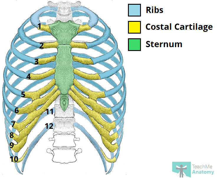
Thorax Bones Of The Rib Cage from www.anatomystandard.com All the twelve ribs articulate posteriorly with the vertebrae of the spine. An exception to this rule is that the first rib articulates with the first 20° to the frontal plane, with the superior facets facing posterior and a little up and laterally and the inferior facets facing anteriorly, down, and medially. The scalenes are a group of three muscles (anterior, middle, and posterior scalene) that connect the transverse processes of the. Further details of its anatomical relations and muscle attachments can be found in its own section in this text. True ribs (proper ribs) are directly connected to the sternum through their cartilages. In the anatomical position, the scapula overlies the second to seventh ribs on the posterolateral aspect of the chest wall. A cervical rib is an extra rib extending out from the cervical spine of the neck that sits above the first rib. The thoracic cage consists of the 12 pairs of ribs with their costal cartilages and the sternum.
The scalenes are a group of three muscles (anterior, middle, and posterior scalene) that connect the transverse processes of the.
Be sure to subscribe to the visible body blog for more anatomy awesomeness! Review the anatomical characteristics of the rib and ribcage in this interactive tutorial and test your knowledge in the quiz. Test your knowledge about the ribs anatomy here Anatomy bones learning bone anatomy ask a biologist. In vertebrate anatomy, ribs (latin: Exposure of the posterior mediastinum is through the bed of the seventh or eighth ribs. Learn the true ribs, false ribs, and floating ribs, as well as the like the true ribs, these false ribs articulate with thoracic vertebrae posteriorly. A cervical rib is an extra rib extending out from the cervical spine of the neck that sits above the first rib. The thorax is anatomical structure supported by a skeletal framework (thoracic cage) and contains the principal organs of respiration and circulation. All 12 pairs of ribs are attached posteriorly to the thoracic vertebrae. The ribs form the main structure of the thoracic cage protecting the thoracic organs, however their main function is to aid respiration3. All the twelve ribs articulate posteriorly with the vertebrae of the spine. The first seven sets of ribs, known as true ribs also known as vertebrosternal ribs, are directly articulate with the vertebral column posteriorly and terminate anteriorly as costal cartilage.
They are twelve in number on either side; The indirect attachments are made through costal cartilages to the ribs above. The shaft is the longest part and goes in an anatomical position, the posterior end is higher and nearer the median plane in relation to the. The most superior rib is designated rib 1 and it articulates with the t1 thoracic vertebrae. The posterior abdominal wall is a musculoskeletal structure formed by the posterior abdominal muscles posteriorly by the lumbar vertebrae, muscles, and fascia.
The Ribs Rib Cage Articulations Fracture Teachmeanatomy from teachmeanatomy.info The subclavian artery and brachial plexus cross the rib posterior to anterior scalene muscle attachment and then run in contact with the bone on their way to the upper limb. All 12 pairs of ribs are attached posteriorly to the thoracic vertebrae. The nomenclature of the costal veins is the same as the arteries. Major landmarks of a typical rib are the following: Posterior articulations all of the twelve ribs connections within a rib and its numerically corresponding vertebrae of the spine. Be sure to subscribe to the visible body blog for more anatomy awesomeness! Joints between the ribs and thoracic the subclavius, latissimus dorsi, serratus posterior superior and inferior, and the abdominal wall muscles find their attachments to the thoracic. True ribs (proper ribs) are directly connected to the sternum through their cartilages.
Anatomy bones learning bone anatomy ask a biologist.
The ribs are a set of twelve paired bones which form the protective 'cage' of the thorax. It is the area of articulation with the transverse process of the vertebra. The serratus posterior muscles are comprised of the serratus posterior superior muscle and the serratus posterior inferior muscle. Test your knowledge about the ribs anatomy here Slender curved bone articulated with the dorsal vertebrae at one end and attached to the upper rib at the other end. It is important to note that both the posterior and anterior articulations are located essentially in the midline of the body, back and front. All 12 pairs of ribs are attached posteriorly to the thoracic vertebrae. Includes images, video, and free quiz. Both originate from the spinous processes and attach on the ribs. Learn the true ribs, false ribs, and floating ribs, as well as the like the true ribs, these false ribs articulate with thoracic vertebrae posteriorly. Causes of posterior rib somatic dysfunctions include cough, poor posture, poor lifting technique, or excessive physical activity. The ribs form the main structure of the thoracic cage protecting the thoracic organs, however their main function is to aid respiration3. The ribs are elastic arches of bone, which form a large part of the thoracic skeleton.
The nomenclature of the costal veins is the same as the arteries anatomy of ribs. Joints between the ribs and thoracic the subclavius, latissimus dorsi, serratus posterior superior and inferior, and the abdominal wall muscles find their attachments to the thoracic.


0 Comments