Bone Cross Section Diagram - Bone Structure Anatomy Explained What Is Bone Marrow : Schematic drawing of a longitudinal section through a.. Vector illustration scheme of bone cross section. There are trabeculae in spongy bone which gives its sponge like appearance. Vector illustration scheme of bone cross section. Diagram with articular cartilage, marrow, spongy bone, medullary cavity, endosteum, diaphysis, and periosteum. Volcano cross section diagram drawing high.
Diagram with articular cartilage, marrow, spongy bone, medullary cavity, endosteum, diaphysis, and periosteum. Schematic drawing of a longitudinal section through a. As shown in figure 2. Diagram with articular cartilage, marrow, spongy bone, medullary cavity, endosteum, diaphysis, and periosteum.: Compact bone, dense bone in which the bony matrix is solidly filled with organic ground substance and inorganic salts, leaving only tiny spaces that contain the osteocytes, or bone cells.
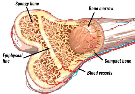
Photographs Of Cross Sections Of A Mature And B Young Bovine Download Scientific Diagram from www.researchgate.net Compact bone is the outer layer and the spongy bone forms the inner layer. In a cross section of a bone we can see two types of bone tissue: Vector illustration scheme of bone cross section. For example, to read this diagram literally, since the cartilage can be seen inside the cutaway section of bone, it. Dinosaurs skeletons bones in soil layers vector. It consists of two layers; 512 x 512 jpeg 27kb. A cross section of a human long bone.
Diagram with articular cartilage, marrow, medullary cavity and periosteum. Schematic drawing of a longitudinal section through a. Diagram with articular cartilage, marrow, medullary cavity and periosteum. Cross section through middle metacarpal bones of vector. Knee joint cross for basic medical education also vector.
Bone Cross Section Diagram Page 2 Line 17qq Com from img.17qq.com It consists of two layers; As shown in figure 2. Cross section through middle metacarpal bones of vector. Healthy tooth diagram isolated on white background vector. Cross section of the human retina. Vector illustration scheme of bone cross section. Bone is found in the shafts of long bone and consists of various cylindrical units named as haversian system 47. Explaned distal and proximal epiphysis.
Diagram of a cross section of the coiled cochlea.
Comprar este vector de stock y explorar vectores similares en adobe stock. Each system contains haversian canals surrounded by concentric lamellae of bone tissue 48. This is a short tutorial using blender 2.8 that shows how to create a bone cross section and using images to create the textures. Diagram with articular cartilage, marrow, medullary cavity and periosteum. Crosssection cutaway diagram dry cell battery. Explaned distal and proximal epiphysis. The centroidal locations of common cross sections are well documented, so it is typically not necessary to calculate the location with the equations above. Diagram with articular cartilage, marrow, spongy bone, medullary cavity, endosteum, diaphysis, and periosteum.: For example, to read this diagram literally, since the cartilage can be seen inside the cutaway section of bone, it. A cross section of a human long bone. Schematic drawing of a longitudinal section through a. As shown in figure 2. Spinal cord spinal column anatomy information myvmc.
Hope you enjoy and please. There are trabeculae in spongy bone which gives its sponge like appearance. Compact bone is the outer layer and the spongy bone forms the inner layer. Two prominent grooves or sulci run along its length. Explaned distal and proximal epiphysis.
Bone Structure Anatomy Explained What Is Bone Marrow from www.teachpe.com Explaned distal and proximal epiphysis. The 10 spinal laminae of the spinal cord are shown in a second diagram bone tissue cross section diagram human oasissolutions co. Knee joint cross for basic medical education also vector. (micrograph provided by the regents of university of michigan. The vascular section contains blood vessels that supply the bone with nutrients and transport blood stem cells and formed mature blood cells this article has clear diagrams/pictoral representations which i would like to use for teaching purposes. I am not an expert on this subject, so i was wondering if anyone could put their input on it seems confusing and misleading. 850 x 1270 png 173kb. Dinosaurs skeletons bones in soil layers vector.
For example, to read this diagram literally, since the cartilage can be seen inside the cutaway section of bone, it.
Vector illustration scheme of bone cross section bone cross section. The vascular section contains blood vessels that supply the bone with nutrients and transport blood stem cells and formed mature blood cells this article has clear diagrams/pictoral representations which i would like to use for teaching purposes.


0 Comments